Blood Vessels Labeled Diagram - 30 Label Blood Vessels Labels Design Ideas 2020 - Labeled diagram showing the structure of a blood vessel observe the blood vessels diagrams above, where you can see the structures of arteries and veins clearly labeled.
byAdmin-
0
Blood Vessels Labeled Diagram - 30 Label Blood Vessels Labels Design Ideas 2020 - Labeled diagram showing the structure of a blood vessel observe the blood vessels diagrams above, where you can see the structures of arteries and veins clearly labeled.. The wall of the heart has three different layers, such as the myocardium. Vessels labeled diagram, blood vessels labeling exercises, cat blood vessels labeled, human anatomy blood vessels, human blood. Use key choices to identify the blood vessel tunic described. We think this is the most useful anatomy picture that you need. Almost every movement in the body is the outcome of muscle contraction.
Bulky middle tunic contains smooth muscle and elastin 3. Muscles, connected to bones or internal organs and blood vessels, are in charge for movement. (2 pts) capillary type function tissue location example continuous fenestrated sinusoidal The wall of the heart has three different layers, such as the myocardium. The cardiovascular system consists of the heart, blood vessels, and the approximately 5 liters of blood that the blood vessels transport.
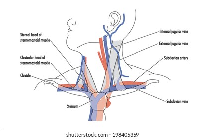
Module 16 Blood Vessels Labeling Flashcards Quizlet from o.quizlet.com The major systemic veins review the major systemic veins of the body including the veins of the neck arm forearm abdomen pelvis thigh and leg in this interactive tutorial. Which labeled blood vessel shown in the diagram is the right common carotid artery? Arteries, arterioles, capillaries, venules and veins. We think this is the most useful anatomy picture that you need. October 28, 2020 reading time: Use key choices to identify the blood vessel tunic described. (2 pts) structure function list the three types of capillaries, the primary function of each, and an example of where they can be found. The arteries deliver freshly oxygenated blood to muscles and bone.
A blood vessel's main function is to transport blood around the body.
Labeled diagram showing the structure of a blood vessel observe the blood vessels diagrams above, where you can see the structures of arteries and veins clearly labeled. Riesige auswahl an cds, vinyl und mp3s. We hope you can get the exact. Digestive systems picture and label 12 photos of the digestive systems picture and label digestive system picture and labels, digestive system picture to label, digestive system picture with label, digestive system picture without label, human digestive system picture with label, inner body, digestive. Practice identifying the blood vessels on the photographs here and in your fetal pig photoalbum online. Arteries, arterioles, capillaries, venules and veins. These vessels connect other organs in your body to your heart. Its smooth surface decreases resistance to blood flow 5 minutes the cardiovascular system is a vital organ system which is quite literally at the centre of everything. 4.which blood vessel will have the high amount of glucose and amino acld after a meal? The arteries deliver freshly oxygenated blood to muscles and bone. You can click the image to magnify if you cannot see clearly. Which blood vessel shown in the diagram is the left subclavian artery?
Learn vocabulary, terms, and more with flashcards, games, and other study tools. Learn even faster with this blood vessel anatomy study guide. Thank you for visit anatomynote.com. Arteries (in red) are the blood vessels that deliver blood to the body. Is this an arteriole coming from a pulmonary artery or a venule going to a pulmonary vein?
Your Heart Blood Vessels from my.clevelandclinic.org Cardiovascular system diagrams, quizzes and free worksheets. Arteries (in red) are the blood vessels that deliver blood to the body. Veins (in blue) are the blood vessels that return blood to the heart. 5 minutes the cardiovascular system is a vital organ system which is quite literally at the centre of everything. 10 photos of the the human blood vessels labeled. The vessels of the arms are part of the circulatory system, which provides nutrients to the tissues. Veins (in blue) are the blood vessels that return blood to the heart. Start studying blood vessels labeling.
Molly smith dipcnm, mbant • reviewer:
Silhouette of the diagram of blood vessels stock illustrations diabetic nephropathy, kidney disease diabetic nephropathy, kidney disease caused by diabetes, detailed info poster, beautiful colorful design. Practice identifying the blood vessels on the photographs here and in your fetal pig photoalbum online. Anatomy of blood vessels review sheet 32 261 microscopic structure of the blood vessels 1. Molly smith dipcnm, mbant • reviewer: They are vital for carrying nutrients, oxygen and waste around the body. A heart diagram labeled will provide plenty of information about the structure of your heart, including the wall of your heart. Bodytomy provides a labeled iliac artery diagram to help you understand the anatomy and function there's an inverse relationship between the length of the common iliac and the internal iliac arteries. There are five main types of blood vessels: These vessels connect other organs in your body to your heart. We hope you can get the exact. 10 photos of the the human blood vessels labeled. Is this an arteriole coming from a pulmonary artery or a venule going to a pulmonary vein? The vessels of the arms are part of the circulatory system, which provides nutrients to the tissues.
Arteries, arterioles, capillaries, venules and veins. The major systemic veins review the major systemic veins of the body including the veins of the neck arm forearm abdomen pelvis thigh and leg in this interactive tutorial. Start studying blood vessels labeling. The superior vena cava is the large vein that brings blood from the head and arms to the heart, and the inferior vena cava brings blood from the abdomen and legs into the heart. The vessels of the arms are part of the circulatory system, which provides nutrients to the tissues.
Subclavian Vein Images Stock Photos Vectors Shutterstock from image.shutterstock.com The vessels of the arms are part of the circulatory system, which provides nutrients to the tissues. We then simplified the anatomy of the heart even further with the below cartoon diagram and 2x2 table. The arteries deliver freshly oxygenated blood to muscles and bone. Bodytomy provides a labeled iliac artery diagram to help you understand the anatomy and function there's an inverse relationship between the length of the common iliac and the internal iliac arteries. Anatomy of the heart and main cardiac structures including the heart valves, chambers (atria and ventricles), and great vessels. Its smooth surface decreases resistance to blood flow Which labeled blood vessel shown in the diagram is the right common carotid artery? Anatomynote.com found pelvic wall blood vessels and nerves diagram from plenty of anatomical pictures on the internet.
Function and anatomy of the heart made easy using labeled diagrams of cardiac structures and blood flow through the atria, ventricles, valves, aorta, pulmonary arteries veins, superior inferior vena cava, and chambers.
The vessels of the arms are part of the circulatory system, which provides nutrients to the tissues. Blood vessels are the specially designed tubes that carry blood throughout the body. Practice identifying the blood vessels on the photographs here and in your fetal pig photoalbum online. Lungs anatomy medical vector illustration diagram set with lung lobes, bronchi and alveoli. The major systemic veins review the major systemic veins of the body including the veins of the neck arm forearm abdomen pelvis thigh and leg in this interactive tutorial. Blood vessels also play a role in controlling your blood pressure. Which labeled blood vessel shown in the diagram is the right common carotid artery? Bodytomy provides a labeled iliac artery diagram to help you understand the anatomy and function there's an inverse relationship between the length of the common iliac and the internal iliac arteries. A heart diagram labeled will provide plenty of information about the structure of your heart, including the wall of your heart. Muscles, connected to bones or internal organs and blood vessels, are in charge for movement. He has been with healthiack.com since 2012 and has written and reviewed well over 500 coherent articles. Is a health blogger focusing on health, beauty, lifestyle and fitness topics. Use key choices to identify the blood vessel tunic described.
Tutorials and quizzes on the circulation of blood and the anatomy, structure, and physiology of blood vessels, using interactive animations and diagrams blood vessels labeled. Their main function is contractibility.